What Is the Intensity Reflection Coefficient in Ultrasound and How Does Acoustic Impedance Affect Measurement and Diagnostic System Performance?
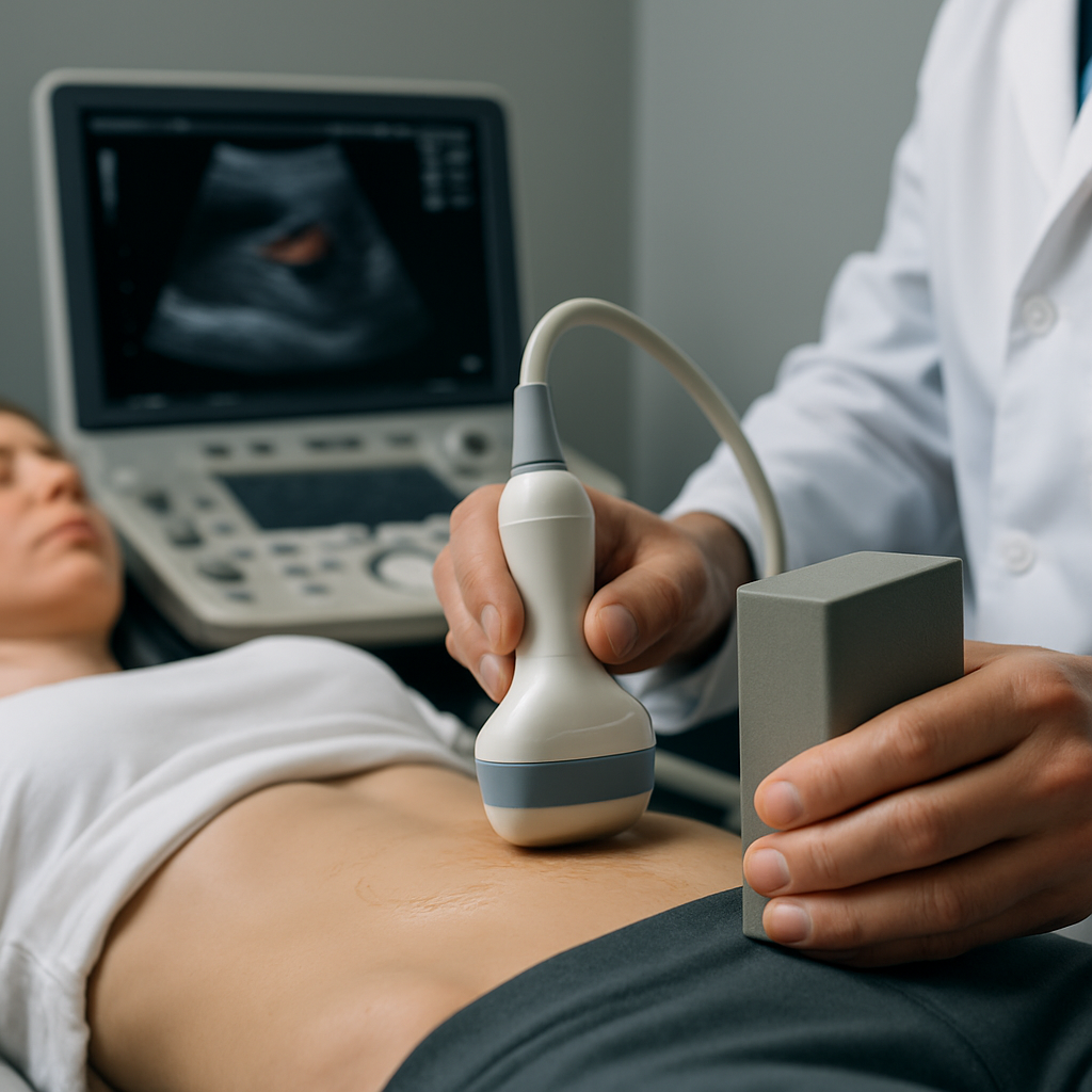
The intensity reflection coefficient in ultrasound describes the fraction of incident ultrasound intensity that is reflected at the boundary between two different media, an essential factor in both diagnostic system performance and accurate measurement of ultrasound parameters. This coefficient, often represented as a ratio or percentage, offers immediate insight into the effectiveness of energy transmission or reflection based on the acoustic impedance mismatch present at tissue or material boundaries. Understanding this fundamental parameter is crucial for anyone working in medical physics, sonography, or related diagnostic imaging fields.
Understanding Intensity Reflection at Acoustic Impedance Boundaries
The reflection of ultrasound at the interface between two media is not random; it depends primarily on a property known as acoustic impedance, defined as the product of a material's density and the speed of sound through it. The greater the difference in acoustic impedance between two media, the greater the proportion of ultrasound reflected, reducing the amount transmitted through the boundary.
This behavior is described mathematically by the equation: IR / I₀ = [(Z₁ – Z₂) / (Z₁ + Z₂)]², where IR is the reflected intensity, I₀ is the incident intensity, and Z₁ and Z₂ are the acoustic impedances of the respective media. As discussed in detail on Sparkl's revision notes, this formula allows precise calculation of how much ultrasound is reflected versus transmitted at boundaries, such as between soft tissue and bone.
Clinically, this means that tissues with similar impedance (like different soft tissues) reflect very little energy, facilitating better ultrasound image penetration, while high-contrast interfaces (such as tissue-bone or tissue-air) can reflect nearly all incident energy, appearing as bright echoes on a scan.
By understanding these concepts, medical professionals can optimize transducer placement and ultrasound frequency selection for maximum diagnostic clarity and system efficiency.
Measurement Parameters and Diagnostic System Considerations
Accurately measuring the intensity reflection coefficient requires careful control of numerous ultrasound parameters, including frequency, spatial average, temporal peak, and power output. Each of these factors can subtly influence the observed coefficient in both laboratory and clinical settings.
For instance, low-intensity pulsed ultrasound often produces a lower spatial average of reflected intensity compared to continuous high-power scanning, translating to safer and more targeted diagnostic applications. Meanwhile, systems that offer high-resolution imaging typically use higher frequencies, which can be more sensitive to impedance mismatches and result in more pronounced reflections at certain boundaries. This helps improve the spatial resolution but may limit penetration depth, especially in denser tissues.
In modern b-ultrasound diagnostic systems, the adjustment of parameters such as the pulse repetition frequency (PRF), dynamic range, and Doppler settings allows clinicians to fine-tune reflection coefficient sensitivity for various clinical uses, whether measuring attenuation in tissues or detecting blood flow anomalies. A comprehensive overview on these measurement strategies can be explored in open-access educational resources such as the LibreTexts Physics of Hearing chapter.
The Role of Acoustic Impedance in Reflection Coefficient Values
Acoustic impedance emerges as the single most influential factor governing the magnitude of the intensity reflection coefficient. In practical ultrasound imaging, the difference between the acoustic impedances of tissues such as muscle, fat, and bone determines their visibility and the clarity of interface boundaries on the diagnostic image.
Smaller differences result in lower reflection coefficients, meaning most ultrasound energy passes through—ideal for visualizing internal organs and tissues. In contrast, a significant impedance mismatch (such as at an air-tissue or bone-tissue boundary) can cause almost total reflection, limiting visualization past that boundary but providing distinct high-contrast indications.
Solidifying comprehension of this association is key for students and practitioners seeking to manipulate scan parameters or interpret reflection coefficients for advanced diagnostic purposes.
Additionally, adjustments in frequency or system power interact with impedance characteristics to further alter measurement outcomes, necessitating a holistic understanding for high-stakes clinical scenarios.
Advanced Applications: Frequency, Power, and Dynamic Range
Modern ultrasound systems not only measure reflected intensities at variable boundaries, but also employ advanced methods like temporal frequency and high spatial resolution modes to optimize diagnostic output. Utilizing high-frequency, low-intensity pulsed scanning in b-mode imaging, for example, enables practitioners to differentiate between subtle tissue boundaries that might share similar acoustic impedance values.
Dynamic range settings within these systems offer yet another level of control, enabling the system to discriminate between low and high reflection coefficients. This improves the ability to capture weak but diagnostically essential echoes, often missed in default operational modes.
Furthermore, power settings are closely monitored to prevent excessive energy reflection that could potentially cause tissue heating or artifacts in high-PRF Doppler studies. This integration of acoustic impedance, measurement parameters, and system configuration allows for safe and effective diagnostic imaging across a wide spectrum of clinical presentations.
Mastering these concepts supports improved patient outcomes and more precise interpretations during both routine and highly specialized sonographic investigations.
The Relationship Between Attenuation, Density, and Reflection Coefficient Behavior
Attenuation, or the gradual loss of ultrasound intensity as it travels through tissue, acts in concert with the reflection coefficient to shape the final detected signal. Densities of different body tissues influence both attenuation and acoustic impedance, meaning that tissues with higher density not only reflect more sound but can also more quickly attenuate ultrasound energy, affecting spatial average and temporal peak intensity measurements.
Practically, understanding how power, attenuation, and reflection coefficients interact enables clinicians to balance system settings for optimal penetration and minimal artifact. For example, lower power and attenuation produce clearer scans in tissues with moderate impedance contrast, while very dense or attenuating tissues may require recalibration or lower diagnostic frequencies to achieve interpretable results.
This complex interplay underscores the significance of integrating reflection coefficient calculations with broader ultrasound physics principles, particularly in ultra high-frequency imaging and specialized applications such as Doppler or b-mode ultrasound diagnostics.
Altogether, these insights equip practitioners and students with the knowledge to excel in clinical diagnostics, system configuration, and physics-based troubleshooting within ultrasound science.
Understanding the intensity reflection coefficient in ultrasound, along with its interdependencies with acoustic impedance, measurement parameters, frequency, and power settings, is essential for optimal performance and confident interpretation in modern b-ultrasound diagnostic systems.


Comments
Post a Comment